Case

0.0The average rating is 0 stars out of 5.

Immediate Implant Placement and Restoration of a Maxillary Left Central Incisor with a Provisional Crown

Leon Pariente, Karim Dada

Leon Pariente and Karim Dada outline the Immediate Implant Placement and Restoration of a Maxillary Left Central Incisor with a Provisional Crown, demonstrating how the hard and soft tissue architecture can be maintained with a Type 1A treatment protocol and a fixed provisional, providing an esthetic and comfortable immediate solution.

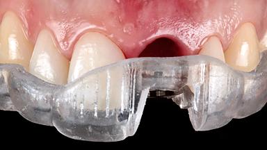
A 34-year-old female patient with high esthetic expectations, a non-smoker with no relevant medical history (ASA class 1), was referred to the practice for replacement of her failing maxillary left central incisor (tooth 21), which exhibited a vertical root fracture as diagnosed by the referring endodontist under the microscope. This case illustrates a type 1A treatment protocol (Gallucci and coworkers 2018) complete with diagnosis, treatment decisions, and clinical and laboratory steps.

Surgical classification
Complex
Prosthodontic classification
Advanced
Source
Treatment Guide 14
Purchase price
10 Academy Coins
CPD/CME
0.25 hours

General Risk Assessment

Patient-related Factors
Smoking Habit None
Oral hygiene Good
Compliance Good
Patient's Expectations High but achievable
Patient-medical Factors
Medical Status Healthy, uneventful healing
Medical Fitness Healthy, able to undergo planned anesthesia and surgical procedure (ASA I)
Medications No medications that would negatively affect the surgical procedure and outcomes.
Radiation Treatment None
Growth Status Complete
Site-related Factors
Periodontal Status No history of periodontal disease, or any active periodontal disease.
Access Adequate
Pathology near the implant site None
Previous surgeries in planned implant site No previous procedures.

Surgical Classification

Surgical Complexity
Timing of placement Immediate Placement (extraction sockets) (Type I)
Simultaneous or Staged grafting procedures Implant placement with simultaneous hard and soft tissue procedures
Anatomy
Bone Volume - Horizontal Adequate
Bone Volume - Vertical Adequate
Keratinized Tissue Sufficient (>4 mm)
Soft Tissue Quality No scars or inflammation
Proximity to vital anatomic structures Minimal risk of involvement
Adjacent Teeth
Papilla Complete
Recession Absent
Interproximal attachment At CEJ
Extractions
Radicular morphology Uniradicular
Available apical bone to achieve primary stability Sufficient height ( ≥ 4 mm) and width (> 2 mm around apex of planned implant)
Socket walls Intact
Thickness of buccal wall 2mm or greater
Anticipated residual defect after implant placement 2 mm or less

Prosthodontic Classification

Complicating Factors
Biological Screw-retained restorations with appropriate contours
Mechanical/Technical Absence of contributing factors
Prosthesis Factors
Prosthetic volume Adequate. Space available for ideal anatomy of the restoration
Inter-occlusal space Adequate. Capable to create an anatomically & functionally correct planned restoration
Volume and characteristics of the edentulous ridge (fixed) Adequate. No adjunctive therapy or prosthetic soft tissue replacement will be necessary
Esthetic Factors
Gingival display at full smile Medium
Shape of tooth crowns Rectangular
Restorative status of neighboring teeth Virgin
Gingival Phenotype Low-scalloped, thick
Bone level on adjacent teeth 5.5 to 6.5 mm to contact point
Occlusal Factors
Occlusal scheme User-defined occlusal scheme achievable
Involvement in occlusion Minimal or no involvement
Occlusal parafunction Absent
Complexity
Loading Protocol Immediate
Implant-supported provisional restoration Required, elevated esthetic and/or functional demands
Timing of placement Immediate Placement (extraction sockets) (Type I)

Esthetic Risk Assessment

Esthetic Risk Assessment
Medical Status Healthy, uneventful healing
Smoking Habit None
Gingival display at full smile Medium
Width of edentulous span 1 tooth (≥ 7mm, standard diameter implant) 1 Tooth (≥ 6mm, narrow diameter implant)
Shape of tooth crowns Rectangular
Restorative status of neighboring teeth Virgin
Gingival Phenotype Low-scalloped, thick
Infection at implant site None
Bone level on adjacent teeth 5.5 to 6.5 mm to contact point
Thickness of buccal wall 2mm or greater
Patient's Expectations High but achievable
Publication date: May 27, 2024 Last review date: May 20, 2024 Next review date: May 20, 2027

ITI QR code Mvc

Share this page

Download the QR code with a link to this page and use it in your presentations or share it on social media.

Download QR code
QR code